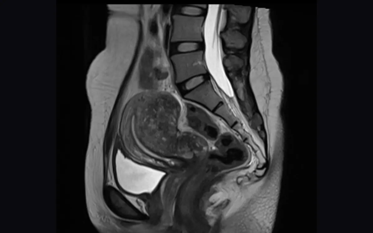
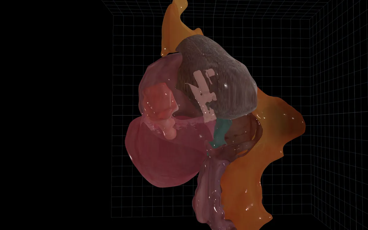
XENA turns pelvic MRI into a patient-specific 3D surgical map, revealing disease extent, organ involvement and critical anatomy so teams can plan the approach, procedure and specialists before the OR.
Clinical evaluations underway with US programs · Investigational · 510(k) in preparation
Today these answers are assembled from the radiology report and the 2D slices, one at a time. XENA holds all four at once.
"…extending to the left uterosacral ligament, with tethering of the anterior rectal wall. Pouch of Douglas obliterated."
Findings described in words.

Reviewed slice by slice.

Disease, organs and critical anatomy in one traceable view.
One patient-specific model, carried from imaging into the OR.
Findings from the pelvic MRI are mapped against the bowel, bladder, ureters, uterus and ovaries. Isolate structures, adjust opacity and return every mapped finding to its source image.
Read complexity from the number of lesions, how deep each one runs and which organs it crosses. Decide the route and the extent of excision before the day of surgery.
Choose a port of entry and preview the laparoscopic view from the patient’s own anatomy. Explore the intended approach before the case.
Open the model, source MRI and saved planning views on iPad during the procedure. No additional hardware.
Suspected bowel, bladder or ureter involvement changes the procedure and who needs to be in the room. XENA shows it while the schedule can still change.
Before consent, walk the patient through her own anatomy, the proposed approach and the specialists who may be involved. The same model the surgical team planned from.
A clearer conversation, from the same source imaging.
Prospective observational pilot · 20 consecutive laparoscopic surgeries for suspected or confirmed moderate-to-severe endometriosis. Thott K, Godbole D. JMIG. 2025;32(11 Suppl):S48. doi:10.1016/j.jmig.2025.09.044.
Works with the MRI the patient already has. Protocol agnostic, 1 to 5 mm slices.
PACS export, PowerShare or secure transfer.
Checked against the radiology read, then reviewed by a practicing surgeon.
MRI and model reviewed side by side. Planning views saved.
The model, source MRI and saved views remain available during the procedure.
Nothing is lost between plan and claim. XENA reads the operative video against the preoperative model and documents work beyond the standard procedure.
Illustrative example of XENA operative-video analysis. Coding decisions remain with the site and its compliance team.
See the mapped disease and its organ relations, and the structures to protect around it.
Flag cases that may require colorectal or urology support before scheduling.
Anticipate staffing, room time and resource needs from the expected procedure.
A preoperative record of disease extent and organ involvement.
3D anatomical segmentation for preoperative planning under HOPPS. Use and payment depend on setting and workflow.
Modifiers 22, 59 / XS and 62 / 66 apply to the operation, not to the model. XENA may support the preoperative record. The operative note must document the work performed.
Eligibility and payment depend on the setting, payer, documented work and applicable policy. Each site should confirm its pathway with its coding and compliance team. XENA is investigational and not yet cleared for clinical use.
Thomassin-Naggara et al., JAMA Network Open 2023, n=605. Independent disease-complexity data, not XENA performance or a demonstrated XENA effect.
Map extent and organ involvement.
Plan excision, approach and team.
Map disease within the uterine wall.
Support treatment and uterine-sparing decisions.
Map number, depth and position.
Plan route, incision, removal and repair.
Map tumor boundaries and critical anatomy.
Plan resection and multidisciplinary care.
The first surgery should be the best surgery.
XENA began with Czuee’s own endometriosis surgeries and one question: why was so much understood only after the operation had begun?
Surgery begins with rich imaging. But when the operation starts, much of that information stays behind. Tissue moves. Anatomy is obscured. The surgeon must decide where to cut and what to protect in real time.
We are building intelligence for the entire operation. Before it, inside it, after it.
Know what the operation will demand. The patient’s MRI becomes a 3D view of the disease and everything it touches. The approach, the procedure and the team are decided before the incision, not after it.
Keep the critical anatomy in view. The next frontier is guidance that holds the plan inside the changing surgical field. What must be removed. What must be protected. The safest path between them.
Turn the operation into evidence. The video is compared with the plan. What was found and what was done become a record that closes this case and improves the next.
CTO & Co-Founder
Stanford Biodesign, previously J&J Medical. Leads imaging AI and medical-device engineering.
COO & Co-Founder
Three-time medtech founder and physicist. Leads hospital adoption, operations and US go-to-market.
Chief Medical Officer
Chief of OB/GYN and practicing MIGS surgeon. Operates weekly and reviews every XENA model.
You choose the cases. We return clinically reviewed models the same week and review them with you.
Moderate-to-severe endometriosis and complex benign gynecology.
Send de-identified DICOM. Receive clinically reviewed models the same week.
Compare each model with the MRI read and available operative findings.
Leave your email and we will schedule a session around one of your cases.
We use your details only to arrange this conversation. We do not share them.